
Tumour
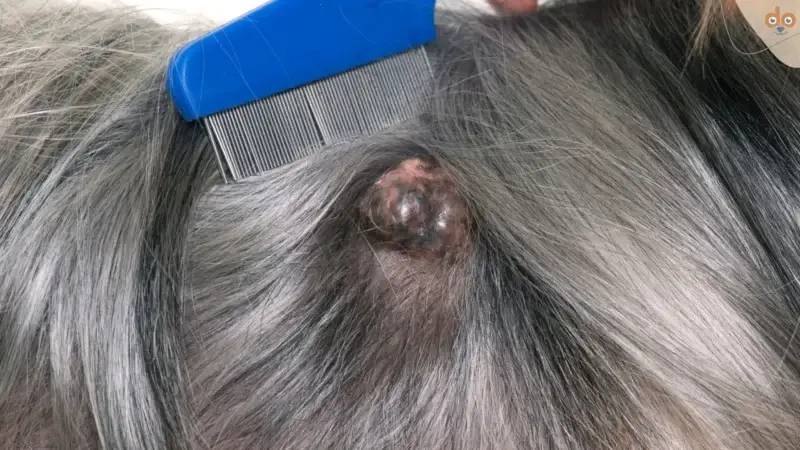
A tumour in a dog is an uncontrolled cell proliferation that can fundamentally occur in any organ.
Content
A dog tumour is an uncontrolled cell proliferation that can fundamentally occur in any organ. Unlike normal cells, which grow and die in a controlled manner, tumour cells multiply without the body’s usual growth brakes.
What distinguishes benign from malignant tumours?
Benign tumours grow slowly, remain locally confined and do not spread to other body regions. Nevertheless, they can cause problems if they press on nerves or organs. Typical examples are lipomas (fatty lumps) or adenomas.
Malignant tumours (cancer) grow aggressively, invade surrounding tissue and can metastasise via the blood or lymphatic vessels. Mast cell tumours, lymphomas or haemangiosarcomas belong in this category.
Which dog breeds have the highest tumour risk?
Certain breeds show significantly increased risks for specific types of tumours:
Golden Retrievers develop lymphomas up to 6 times more frequently than other breeds. Boxers have an increased risk of mast cell tumours; approximately 20% of all skin tumours in Boxers are mast cell tumours. German Shepherds tend to develop haemangiosarcomas, particularly in the spleen.
In large breeds such as Rottweilers or Dobermans, osteosarcoma (bone cancer) occurs approximately 200 times more frequently than in small dogs. The risk increases drastically from a body weight of 40 kg.
How can I recognise a tumour in my dog?
Most tumours initially appear as palpable nodules under the skin. These can be hard or soft, mobile or firmly attached. You should regularly palpate your dog, particularly the armpits, groin and neck.
Further warning signs include non-healing wounds, persistent lameness without an apparent cause, unexplained weight loss despite normal food intake or chronic breathing difficulties. In older dogs from 8 years onwards, the risk increases significantly.
Some tumours only cause symptoms late on. Splenic tumours, for example, often go unnoticed until they rupture and cause internal bleeding.
How does the vet make the diagnosis?
The first step is usually a fine-needle aspiration – cells are taken from the suspicious tissue with a thin needle. This method is low-pain and quickly provides initial indications as to whether it is benign or malignant.
A complete histopathology (tissue examination) is, however, the gold standard. Only it shows the exact tumour type and staging (spread). X-rays, ultrasound or CT scans supplement the diagnostics to detect metastases.
What treatment options are available?
Surgical removal remains the most important therapy for most tumours. It is crucial to remove sufficient healthy tissue along with it; for mast cell tumours, for example, a safety margin of at least 2 cm in all directions.
Chemotherapy in dogs differs from human medicine: the aim is life extension with good quality of life, not healing at any cost. Most dogs tolerate it well and show no severe side effects such as hair loss.
Radiotherapy is particularly suitable for tumours in locations that are difficult to operate on, such as the nose or brain. Modern techniques allow precise irradiation with few side effects.
What is the prognosis for tumours?
For benign tumours, the prognosis after complete removal is excellent. For malignant tumours, it depends heavily on the type and stage. Grade I mast cell tumours have a very good prognosis, whereas Grade III often leads to death quickly.
Lymphomas usually respond well to chemotherapy – 80% of dogs achieve remission. The median survival time is 12–14 months. Osteosarcomas, on the other hand, often have a prognosis of only 4–6 months despite amputation without additional chemotherapy.
More from the Wiki
All wiki entries →Rhubarb
Rhubarb is a knotweed whose leaves are rich in soluble oxalic acid. For dogs, the leaves …
Why is dog faeces white?
White dog faeces occurs when normal digestion is disrupted or certain substances change the colour of …
Dog licks its anus, what to do?
Anal licking in dogs is a normal grooming behaviour that serves for cleaning and emptying the …
Why do some dogs eat their vomit?
Eating one's own vomit is normal behaviour in dogs, stemming from evolutionary instincts.