
Thorax
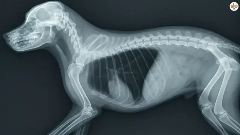
The thorax is the bony protective shield around the heart and lungs of a dog, consisting of 13 pairs of ribs, the sternum and the associated muscles.
Content
In dogs, the thorax refers to the ribcage. It is formed by the spine, ribs, sternum, and associated muscles and connective tissue structures. The ribcage encloses the thoracic cavity and protects vital organs such as the heart and lungs.
Dogs usually have 13 pairs of ribs. The ribs are connected to the thoracic spine via joints. The front ribs connect to the sternum via costal cartilage, while the rear ribs partly end in a common rib arch or terminate freely.
The shape of the thorax varies depending on body build and breed. Deep-chested dogs, for example, have a distinctly differently shaped ribcage than short-legged or compactly built dogs.
Which organs and structures are located in the thorax?
The thoracic cavity contains mainly the lungs and the heart. Additionally, the following pass through the thorax, among others:
- Trachea
- Oesophagus
- major blood vessels
- nerves and lymphatic vessels
The lungs lie in the thoracic cavity and are surrounded by the pleura, or chest membrane. Between the two lung lobes lies the mediastinum, the central area of the thoracic cavity. Located there, among other things, are the heart, major blood vessels, trachea, and oesophagus.
The heart lies within the pericardium (heart sac). Its exact position and orientation depend, among other factors, on body build and breed. A general statement that the dog’s heart is “more central and slightly tilted to the left” describes the anatomy only inadequately.
Which diseases and injuries can affect the thorax?
Diseases of the thorax can affect the airways, lungs, heart, pleura, blood vessels, or the bony structures of the ribcage.
Possible problems include, for example:
- Pneumothorax, i.e. air in the pleural space
- Pleural effusion, i.e. fluid in the pleural space
- Hemothorax, a collection of blood in the chest cavity
- Lung injuries, e.g. after an accident
- Rib fractures
- Lung diseases
- Heart diseases
- Tumours in the chest cavity
- Diaphragmatic hernias
Especially after trauma, multiple structures may be injured simultaneously. Possible consequences include, among others, lung contusions, pneumothorax, bleeding in the chest cavity, arrhythmias, rib fractures, and diaphragmatic hernias.
What is a pneumothorax?
In a pneumothorax, air enters the pleural space, i.e., the space between the lung and the chest wall. As a result, the lung may partially or completely collapse.
A pneumothorax can occur, for example, following an accident or injury to the chest. It can also arise spontaneously if air from the lung tissue enters the pleural space. In a tension pneumothorax, pressure in the thoracic cavity continues to rise, which can significantly impair respiration and circulation.
Possible signs include rapid or laboured breathing, shallow breaths, and weakness. A severe pneumothorax is a medical emergency.
What is a pleural effusion?
In pleural effusion, fluid accumulates in the pleural space. This does not refer to fluid ‘around the lungs’ in the sense of a normal anatomical state, but rather to a pathological accumulation within the thoracic cavity.
Possible causes include, among others, bleeding, inflammation, tumours, or disorders of blood and lymph drainage. Depending on the type of fluid, distinctions are made, for example, between haemothorax (blood), chylothorax (lymph with high fat content), and other forms.
A large pleural effusion can displace the lung, thereby making breathing more difficult. Affected dogs may, for example, breathe rapidly and shallowly, appear weak, or show increased work of breathing.
What is a diaphragmatic hernia?
In a diaphragmatic hernia, abdominal contents pass through an opening or tear in the diaphragm into the thoracic cavity.
Traumatic diaphragmatic hernias in dogs often occur after severe accidents. Congenital forms also exist. Depending on which organs enter the thoracic cavity and how severely they affect the lungs or other structures, symptoms can range from unremarkable to severe respiratory distress.
Diagnosis is frequently made using X-rays. Ultrasound or further imaging techniques may also be employed.
In cases of traumatic diaphragmatic hernia, the dog is initially stabilised. Definitive treatment usually involves surgical closure of the diaphragmatic tear. However, immediate surgery is not always the first measure, as life-threatening circulatory or respiratory problems must be addressed first.
How dangerous are rib fractures?
Ribs can break primarily due to blunt force trauma, for example in road traffic accidents. Individual rib fractures can heal without surgery. However, what is decisive is not only the fracture itself, but also whether the lungs, blood vessels, or other structures have been injured simultaneously.
Multiple broken ribs can lead to an unstable chest wall (flail chest). In this condition, part of the chest wall moves in opposition to normal respiratory movement during breathing. This can significantly impair respiration.
Treatment depends on the extent of the injuries. This includes, in particular, adequate pain management and treatment of other injuries. Not every rib fracture requires surgery. In cases of severe thoracic trauma, intensive care monitoring may be necessary.
How can you recognise problems in the thorax?
Thoracic diseases can manifest differently. Possible signs include:
- rapid or laboured breathing
- shallow breathing
- increased work of breathing
- coughing
- pain when breathing or moving
- reduced exercise tolerance
- weakness
- pale or bluish mucous membranes
- collapse
Rapid breathing after physical exertion or in high temperatures may, however, be a normal reaction. However, an noticeably increased respiratory rate or work of breathing at rest should be investigated by a vet.
Severe respiratory distress, bluish mucous membranes, collapse, or rapid deterioration are emergency signs.
Following a severe accident, a dog should be examined even if only minor external injuries are visible, as internal injuries cannot be reliably assessed based on the skin alone.
How is the thorax examined?
The examination begins with a clinical assessment. This includes, among other things, observing respiration and auscultating the heart and lungs.
Depending on the suspicion, various imaging techniques may be used:
- X-rays of the thorax can provide indications of rib fractures, pneumothorax, pleural effusion, changes in lung tissue or changes in heart size, among other things. However, in the case of thoracic trauma, X-rays are only part of the possible diagnostics.
- Ultrasound: The ultrasound examination can be particularly helpful in diseases of the pleura and fluid accumulations. Certain changes to the heart can also be examined using ultrasound.
- A special form is echocardiography, i.e. cardiac ultrasound. This allows assessment of heart valves, heart chambers, atria and heart function, among other things.
- CT and further procedures: A computed tomography (CT) scan can provide more detailed images of the ribcage and is used, for example, in certain tumours, complex injuries or for surgical planning. An MRI is used significantly less frequently for thoracic diseases.
- In the case of fluid or air in the chest cavity, a thoracentesis can also be performed. In this procedure, fluid or air is removed from the pleural space using a needle or catheter. The fluid obtained can then be examined.
More from the Wiki
All wiki entries →Plum
The plum is a stone fruit of the genus Prunus. Its stone contains cyanogenic glycosides; the …
Rhubarb
Rhubarb is a knotweed whose leaves are rich in soluble oxalic acid. For dogs, the leaves …
Why is dog faeces white?
White dog faeces occurs when normal digestion is disrupted or certain substances change the colour of …
Dog licks its anus, what to do?
Anal licking in dogs is a normal grooming behaviour that serves for cleaning and emptying the …